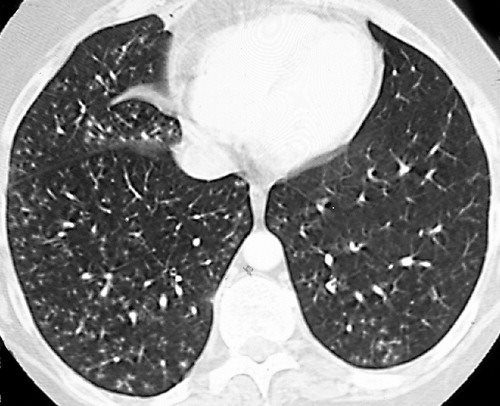
tree in bud opacities in lungs
Multiple causes for tree-in-bud TIB opacities have been reported. Bat wing opacities also known as butterfly opacities refer to a pattern of bilateral perihilar lung shadowing.

Tree In Bud Sign And Bronchiectasis Radiology Case Radiopaedia Org
Bat wing pulmonary opacities can be caused by.
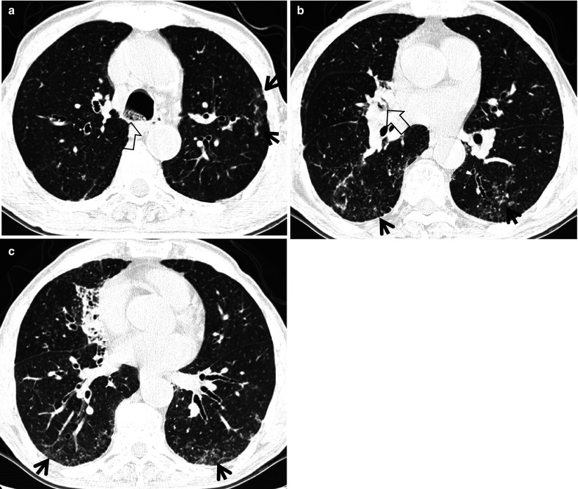
. High-resolution CT scan of the thorax demonstrates central bronchiectasis a hallmark of allergic bronchopulmonary aspergillosis right arrow and the peripheral tree-in-bud appearance of centrilobular opacities left arrow which represent mucoid impaction of the small bronchioles. It is classically described on a frontal chest radiograph but can also refer to appearances on chest CT 34. They are suggestive for the diagnosis of congestive heart failure but are also seen in various non-cardiac conditions such as pulmonary fibrosis interstitial deposition of heavy metal particles or carcinomatosis of the lung.
However to our knowledge the relative frequencies of the causes have not been evaluated. Chronic Kerley B lines may be caused by fibrosis or hemosiderin deposition caused by recurrent pulmonary edema. Welcome to pyradiomics documentation This is an open-source python package for the extraction of Radiomics features from medical imaging.
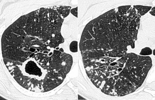
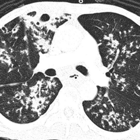
The visceral pleura is the one that covers both the lungs. Pulmonary edema especially cardiogenic pneumonia. In the right mid-lung nodular opacities are in a tree-in-bud distribution suggestive of endobronchial spread.
The pleural space is a cavity between the 2 pleurae into which the lung protrudes. There are widespread TIB opacities throughout both lungs with predominance in the anterior nondependent portions of the lungs a finding that would normally suggest a diagnosis. The parietal and visceral pleurae are separated by a thin layer of fluid.
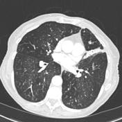
While the tree-in-bud appearance usually represents an endobronchial spread of infection given the proximity of small pulmonary arteries and small airways sharing branching morphology in the bronchovascular bundle a rarer cause of the tree-in-bud sign is infiltration of the small pulmonary arteriesarterioles or axial interstitium 367. TIB opacities represent a normally invisible branches of the bronchiole tree 1 mm in diameter that are severely impacted with mucous pus or fluid with resultant dilatation and budding of the terminal bronchioles 2 mm in diameter 1 photo. Small patchy peripheral opacities are also present in the left lower lobe.
The visceral and parietal pleura are smooth serous membranes continuous with each other at the lung hila. Although initially described in 1993 as a thin-section chest CT finding in active tuberculosis TIB opacities are by. With this package we aim to establish a reference standard for Radiomic Analysis and provide a tested and maintained open-source platform for easy and reproducible Radiomic Feature extraction.
Coronal reconstructed computed tomography image shows the lingular cavity with irregular nodules and right mid-lung nodular opacities in a 43-year-old man who.
Tree In Bud Sign Lung Radiology Reference Article Radiopaedia Org
Tree In Bud Sign Radiology Key
It Is Not Always Tuberculosis Tree In Bud Opacities Leading To A Diagnosis Of Sarcoid Shm Abstracts Society Of Hospital Medicine
Tree In Bud Sign Lungs
2
2

Pdf Tree In Bud

Tree In Bud Pattern Pulmonary Tb Eurorad
View Of Tree In Bud The Southwest Respiratory And Critical Care Chronicles
2
2

Tree In Bud Pattern Pulmonary Tb Eurorad

Tree In Bud Sign Lung Radiology Reference Article Radiopaedia Org

Tree In Bud Sign And Bronchiectasis Radiology Case Radiopaedia Org

Pdf Tree In Bud Semantic Scholar
Tree In Bud Sign Lungs
Tree In Bud Pattern Pulmonary Tb Eurorad

Tree In Bud Pattern Pulmonary Tb Eurorad
2